소화기 (소화기계/소화기관) 구조, Anatomy of digestive system
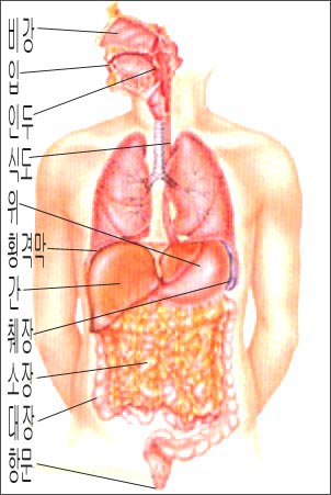
그림 1. 소화기계(소화계) 해부도
출처-Used with permission from Galaxo Wellcome과 소아가정간호 백과
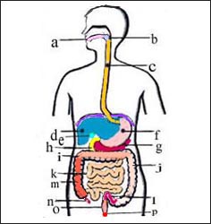
그림 2. 소화기계(소화계) 해부도
a-입, b-인두, c-식도, d-간, e-담낭, f-위, g-췌장, h-십이지장, i-횡행결장, j-하행결장, k-소장, l-S상결장, m-상행결장, n-충수, o-직장, p-항문
Copyright Ⓒ 2013 John Sangwon Lee, MD., FAAP
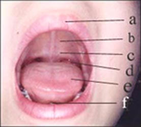
사진 3. 입안의 구조
a-입술, b-경구개, c-연구개, d-편도, e-혀, f-소대
Copyright Ⓒ 2013 John Sangwon Lee, MD., FAAP
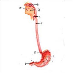
그림 4. 입, 인두, 식도, 위의 해부도
a-입, b-비강, c-인두(인후), d-혀, e-후두, f-식도, g-분문, h-위, i-위벽, j-십이지장, k-유문
출처-소아가정간호 백과와 Used with permission from Galaxo Wellcome
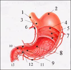
그림 5. 식도, 위, 십이지장
1-식도, 2-위 저, 3-분문, 4-종횡 섬유조직, 5-윤상 섬유조직, 6-혈관, 7-사위 섬유조직, 8-위벽, 9-위 동, 10-십이지장의 상부, 11-혈관, 12-유문, 13-유문 괄약근
출처-소아가정간호 백과와 Used with permission from Galaxo Wellcome
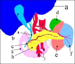
그림 6. 간과 그 부위의 해부도
a-간, b-담낭, c-십이지장 유두(담즙과 췌장액이 분비되는 출구), d-부신, e-좌 신장, f-지라(비장), g-십이지장, h-췌장, i-총수담관, j-혈관
Copyright Ⓒ 2013 John Sangwon Lee, MD., FAAP
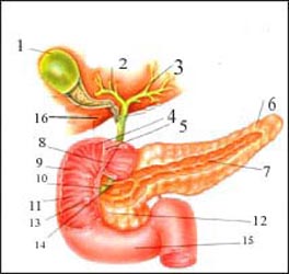
그림 7.간, 담낭, 담도, 췌장, 십이지장
1-담낭, 2-우 간 담도, 3-좌 간 담도, 4-십이지장, 5-유문괄약근, 6-췌장의 미부, 7-췌장 관, 8-유문, 9-십이지장, 10-총수 담관, 11-십이지장, 12-췌장의 두부, 13-십이지장 유두, 14-췌장 관, 15-십이지장, 16-간
출처-소아가정간호 백과와 Used with permission from Galaxo Wellcome
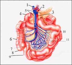
그림 8. 소장과 대장의 혈관
1-문 정맥, 2-비장정맥, 3-대동맥, 4-십이지장, 5-췌장, 6-상행결장, 7-회장동맥, 8-맹장, 9-충수, 10-공장 동맥, 11-장간막 동맥호
출처-소아가정간호 백과와Used with permission from Galaxo Wellcome
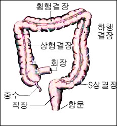
그림 9. 대장(상행결장+횡행결장+하행결장+S상결장)
Copyright Ⓒ 2011 John Sangwon Lee, MD., FAAP
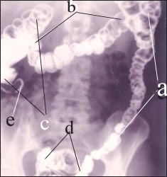
사진 10. 바륨 에네마로(바륨관장)로 본 대장 X-선 사진
a-하행 결장, b-횡행 결장, c-상행 결장, d-S상 결장, e-충수
Copyright Ⓒ 2011 John Sangwon Lee, MD., FAAP
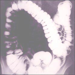
사진 11. 바륨관장으로 본 대장 X-선 사진
Copyright Ⓒ 2011 John Sangwon Lee, MD,, FAAP
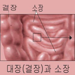
그림 12. 대장관과 소장관
Copyright Ⓒ 2011 John Sangwon Lee, MD., FAAP
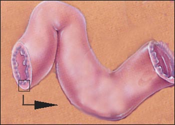
그림 13. 소장관의 일부
Copyright Ⓒ 2011 John Sangwon Lee, MD., FAAP
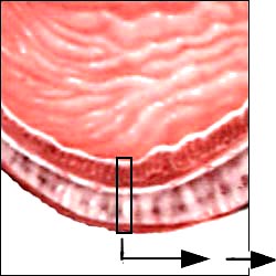
그림 14. 소장관 벽의 일부
Copyright Ⓒ 2011 John Sangwon Lee, MD., FAAP
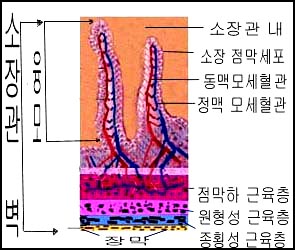
그림 15.현미경으로 본 소장관 벽 조직.
소장관 벽은 점막층+점막층 하 조직+근육층+장막(장막층) 총 4층으로 구성되었다.
Copyright Ⓒ 2011 John Sangwon Lee, MD., FAAP
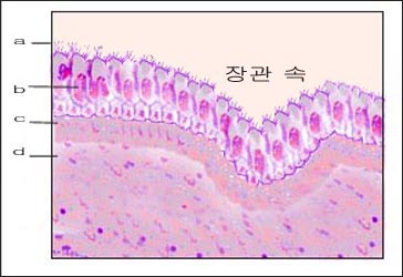
그림 16. 소장관 벽의 점막층 조직
a-소장관 벽 점막층에 있는 융모,
b-소장관 벽 점막층에 있는 점막세포,
c, d-점막층 하 조직
어떤 종류의 경구용 항생제로 감염병을 치료할 때나 바이러스 감염이나 박테리아 감염 등으로 소장관 벽 점막층에 있는 융모나 소장관 벽 점막층에 있는 점막세포 등이 손상될 수 있다.
점막층 세포가 손상되면 그로 인해서 먹은 음식물이 정상적으로 소화되지 않고 정상적으로 흡수되지 않을 수 있다. 또 체내에 있는 체액이나 혈장이 손상된 소장관 벽 점막층을 통해서 장관 속으로 비정상적으로 더 많이 분비될 수 있다. 이렇게 해서 설사를 하게 된다.
Copyright Ⓒ 2011 John Sangwon Lee, MD., FAAP
Anatomy of the digestive system
출처 및 참조 문헌 Sources and references
- NelsonTextbook of Pediatrics 22ND Ed
- The Harriet Lane Handbook 22ND Ed
- Growth and development of the children
- Red Book 32nd Ed 2021-2024
- Neonatal Resuscitation, American Academy Pediatrics
- www.drleepediatrics.com 제1권 소아청소년 응급 의료
- www.drleepediatrics.com 제2권 소아청소년 예방
- www.drleepediatrics.com 제3권 소아청소년 성장 발육 육아
- www.drleepediatrics.com 제4권 모유,모유수유, 이유
- www.drleepediatrics.com 제5권 인공영양, 우유, 이유식, 비타민, 미네랄, 단백질, 탄수화물, 지방
- www.drleepediatrics.com 제6권 신생아 성장 발육 육아 질병
- www.drleepediatrics.com제7권 소아청소년 감염병
- www.drleepediatrics.com제8권 소아청소년 호흡기 질환
- www.drleepediatrics.com제9권 소아청소년 소화기 질환
- www.drleepediatrics.com제10권. 소아청소년 신장 비뇨 생식기 질환
- www.drleepediatrics.com제11권. 소아청소년 심장 혈관계 질환
- www.drleepediatrics.com제12권. 소아청소년 신경 정신 질환, 행동 수면 문제
- www.drleepediatrics.com제13권. 소아청소년 혈액, 림프, 종양 질환
- www.drleepediatrics.com제14권. 소아청소년 내분비, 유전, 염색체, 대사, 희귀병
- www.drleepediatrics.com제15권. 소아청소년 알레르기, 자가 면역질환
- www.drleepediatrics.com제16권. 소아청소년 정형외과 질환
- www.drleepediatrics.com제17권. 소아청소년 피부 질환
- www.drleepediatrics.com제18권. 소아청소년 이비인후(귀 코 인두 후두) 질환
- www.drleepediatrics.com제19권. 소아청소년 안과 (눈)질환
- www.drleepediatrics.com 제20권 소아청소년 이 (치아)질환
- www.drleepediatrics.com 제21권 소아청소년 가정 학교 간호
- www.drleepediatrics.com 제22권 아들 딸 이렇게 사랑해 키우세요
- www.drleepediatrics.com 제23권 사춘기 아이들의 성장 발육 질병
- www.drleepediatrics.com 제24권 소아청소년 성교육
- www.drleepediatrics.com 제25권 임신, 분만, 출산, 신생아 돌보기
- Red book 29th-31st edition 2021
- Nelson Text Book of Pediatrics 19th- 21st Edition
- The Johns Hopkins Hospital, The Harriet Lane Handbook, 22nd edition
- 응급환자관리 정담미디어
- Pediatric Nutritional Handbook American Academy of Pediatrics
- 소아가정간호백과–부모도 반의사가 되어야 한다, 이상원 저
- The pregnancy Bible. By Joan stone, MD. Keith Eddleman, MD
- Neonatology Jeffrey J. Pomerance, C. Joan Richardson
- Preparation for Birth. Beverly Savage and Dianna Smith
- 임신에서 신생아 돌보기까지. 이상원
- Breastfeeding. by Ruth Lawrence and Robert Lawrence
- Sources and references on Growth, Development, Cares, and Diseases of Newborn Infants
- Emergency Medical Service for Children, By Ross Lab. May 1989. p.10
- Emergency care, Harvey Grant and Robert Murray
- Emergency Care Transportation of Sick and Injured American Academy of Orthopaedic Surgeons
- Emergency Pediatrics A Guide to Ambulatory Care, Roger M. Barkin, Peter Rosen
- Quick Reference To Pediatric Emergencies, Delmer J. Pascoe, M.D., Moses Grossman, M.D. with 26 contributors
- Neonatal resuscitation Ameican academy of pediatrics
- Pediatric Nutritional Handbook American Academy of Pediatrics
- Pediatric Resuscitation Pediatric Clinics of North America, Stephen M. Schexnayder, M.D.
-
Pediatric Critical Care, Pediatric Clinics of North America, James P. Orlowski, M.D.
-
Preparation for Birth. Beverly Savage and Dianna Smith
-
Infectious disease of children, Saul Krugman, Samuel L Katz, Ann A.
- 제4권 모유, 모유수유, 이유 참조문헌 및 출처
- 제5권 인공영양, 우유, 이유, 비타민, 단백질, 지방 탄수 화물 참조문헌 및 출처
- 제6권 신생아 성장발육 양호 질병 참조문헌 및 출처
- 소아과학 대한교과서
Copyright ⓒ 2014 John Sangwon Lee, MD., FAAP
“부모도 반의사가 되어야 한다”-내용은 여러분들의 의사로부터 얻은 정보와 진료를 대신할 수 없습니다.
“The information contained in this publication should not be used as a substitute for the medical care and advice of your doctor. There may be variations in treatment that your doctor may recommend based on individual facts and circumstances.
“Parental education is the best medicine.
-
Copyright ⓒ 2014 John Sangwon Lee, MD., FAAP
“부모도 반의사가 되어야 한다”-내용은 여러분들의 의사로부터 얻은 정보와 진료를 대신할 수 없습니다.
“The information contained in this publication should not be used as a substitute for the medical care and advice of your doctor. There may be variations in treatment that your doctor may recommend based on individual facts and circumstances.
“Parental education is the best medicine.